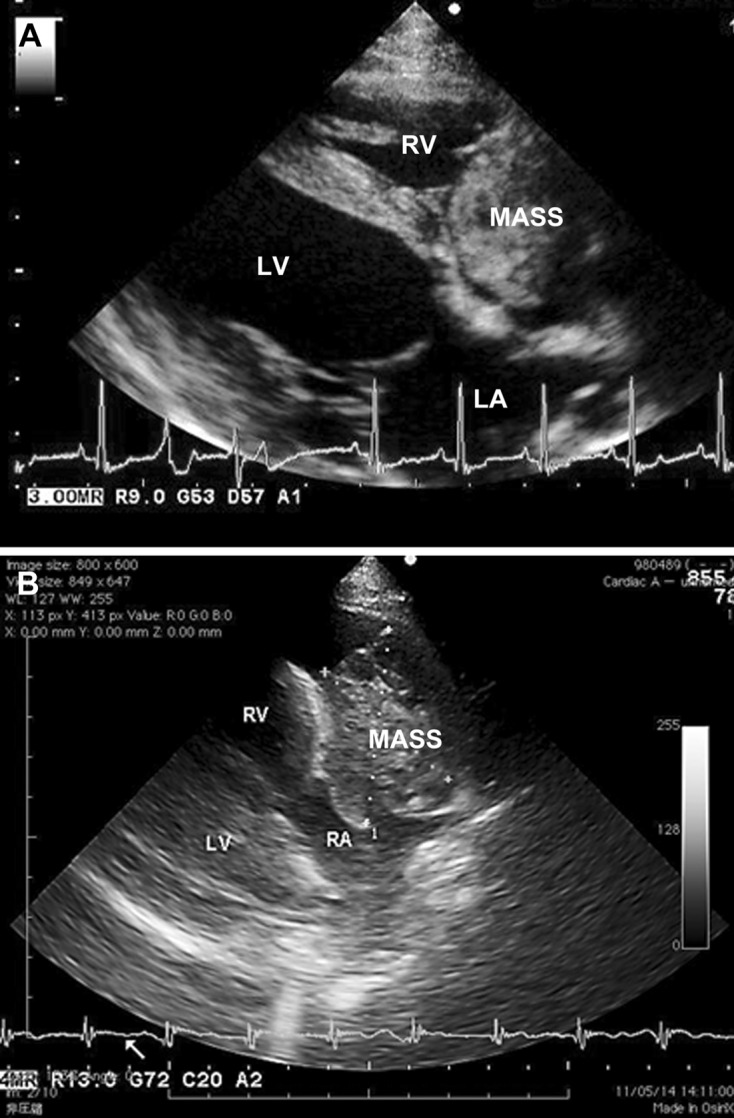
Fig. 2.

Two-dimensional echocardiograms (long-axis view from the right parasternal location) of dogs with primary cardiac hemangiosarcoma, showing a cavitary and cystic mass (MASS) associated with the right auricle (A) and a cavitated soft tissue mass (MASS) occupying the right atrial cavity. LA, left atrium; LV, left ventricle; RA, right atrium, RV, right ventricle.
