Figure 4.
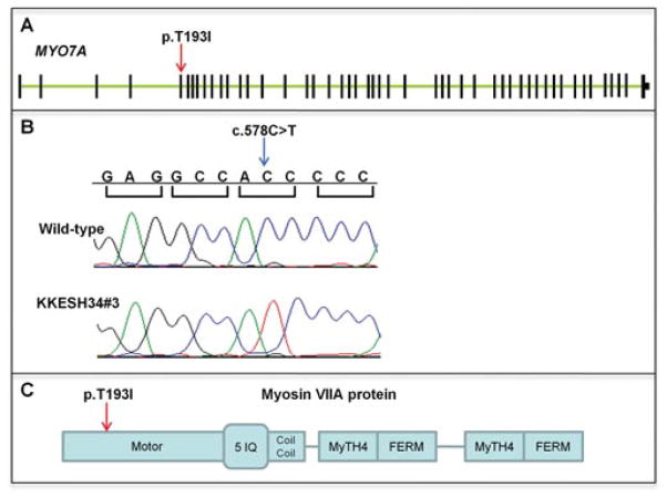
A missense mutation is identified in MYO7A. A: Exon–intron structure of MYO7A. Exons are shown as black boxes. The missense mutation p.T193I is located in the fifth exon (red arrow). B: Sequence traces of control and affected members. A homozygous missense mutation in c.578 is identified in affected member KKESH34#3 (blue arrow). C: Schematic view of the MYO7A protein structure. The p.T193I mutation is located within the motor domain.

