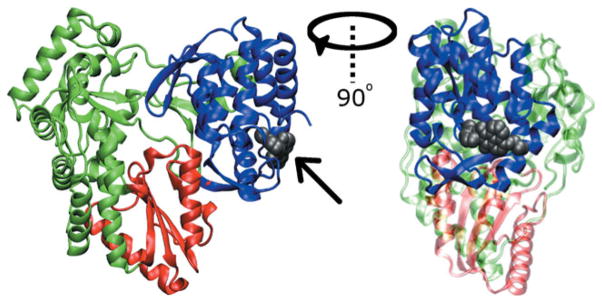
Figure 1.

Cartoon representation of NS5B from PDB structure 2WHO. The fingers domain is shown in green, the palm domain in red, and the thumb domain in blue. Allosteric binding site NNI2 is shown occupied by the ligand VGI (gray space filling representation identified by arrow) from the 2WHO structure. The right panel represents a 90° rotation about the vertical axis relative to the left.
