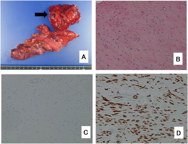
Figure 3.

Resected surgical specimen. (A) The resected tumor was approximately 65 mm × 35 × 55 mm in diameter. (B) Histopathologically, H & E staining showed that the tumor cells had no necrosis and mitotic activity, as shown at × 200. The tumor cells were negative for c-kit, as shown at × 200, (C) and positive for CD34, as shown at × 200, (D) using immunohistochemistry.
