
Figure 2.
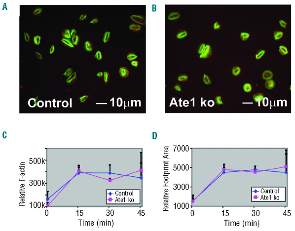
Platelet spreading on fibrinogen in response to thrombin. (A and B) Control and ATE1 knockout (ko) platelets were allowed to spread on fibrinogen-coated coverslips in the presence of thrombin. The cells were fixed and stained with phalloidin to detect F-actin (green) and antibodies were directed to detect β-actin (red). F-actin was concentrated centrally in the cortex of the spread platelets while β-actin was detected mostly at the edges. (C) Relative F-actin levels of platelets were assessed by flow cytometry staining with Alexa 488-Phalloidin. (D) The relative area (footprint size) of spread platelets is shown. The mean ± standard deviation are shown.