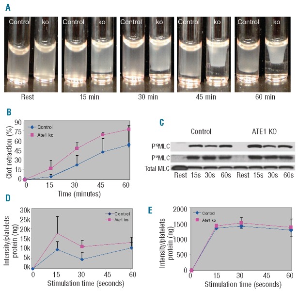
Figure 4.

Increased clot retraction and phosphorylation of myosin in platelets lacking ATE1. (A) Platelet-rich plasma from ATE1 knockout (ko) and control mice was stimulated with thrombin at 37°C to induce clotting. Clot formation and retraction were continuously photographed before and after thrombin stimulation at 15 min intervals. Representative images at each time point show increased clot retraction in ATE1 ko platelets as compared to control platelets. (B) Quantification of clot retraction at each time point was measured as the percentage of the retracted area as compared to the initial total area of the clot (n=5.) Deletion of ATE1 significantly increased clot retraction (P<0.05, except at 60 min). The mean ± standard deviation are shown. (C) Phosphorylation of RLC at residues Ser19 and Thr18 was undetectable in resting platelets, but increased following stimulation with thrombin. Phosphorylation of RLC at either Ser19 (D) or Thr18 (E) was quantified based on the immunoblots of platelets stimulated with thrombin. The level of Ser19-RLC phosphorylation was significantly increased in platelets lacking ATE1 as compared to that in the controls (P<0.05) at 15 seconds (P<0.03) and at 30 seconds (P<0.05) after agonist stimulation. In contrast, Thr18-RLC phosphorylation remained the same in both groups. The mean ± standard deviation are shown.
