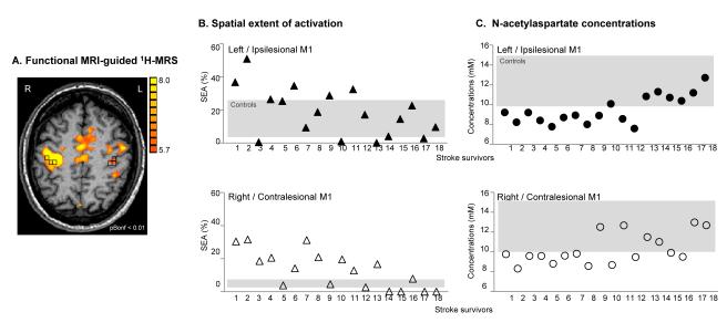
Fig. 1.
Motor-related cortical activation during a handgrip task executed with the impaired right hand in a 58-age old patient who had experienced infarct involving the left striato-capsular area (Patient 18, Table 1). 1H-MRSI slab (grey rectangle) was positioned on axial T1-weigthed MR image at the level of motor-related motor/premotor activation. Spectroscopic voxels (black squares) were selected based on M1, PMd, and SMA activation (or anatomical landmarks in case the activation tended to zero). L=left, R=right

