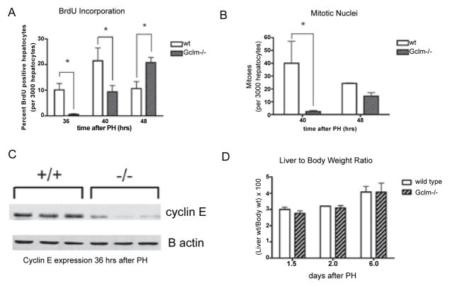
Figure 3.
Delayed hepatocyte proliferation after PH in Gclm−/− mice.
A: BrdU incorporation in hepatocytes after PH, presented as the percentage of positively staining hepatocytes in 3000 cells examined for each mouse. *=p<0.05. BrdU: bromodeoxyuridine; PH: partial hepatectomy; wt: wild type. B: Hepatocyte mitotic counts after PH, presented as number of mitoses per 3000 hepatocytes examined. *=p<0.05. C: Western blot demonstrating cyclin E expression 36 hours after PH in wild type (+/+) and Gclm−/− (−/−) mice.
D: Liver weights expressed as a percentage of body weight 1, 2, and 6 days after PH in wt (wild type) and Gclm−/− mice; n=3–6 mice per genotype per time point.

