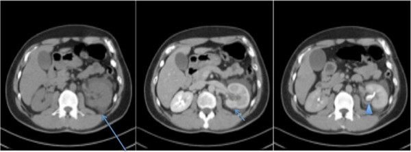
Figure 1.

Axial computed tomography image in the plain, nephrographic and pyelographic phases. A large amount of perinephric fluid is seen on the left side (long arrow in first panel). Delayed enhancement of the left kidney with delayed excretion of contrast is apparent (short arrow in second panel), but no solid mass or urinary calculus is seen. Dependent material is seen in the nondilated collecting system of the left kidney consistent with blood (arrowhead in third panel).
