
Figure 10.
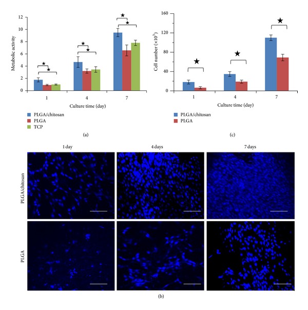
Viability and proliferation of PLGA/chitosan and PLGA nanofibers: (a) metabolic activity of 3T3 fibroblasts on PLGA/chitosan and PLGA nanofibers and TCP measured by an MTS assay, (b) DAPI-stained nuclei of 3T3 fibroblasts on PLGA/chitosan, and PLGA nanofibers after 1, 4, and 7 days; scale bar representing 100 μm, and (c) proliferation of 3T3 fibroblasts on PLGA/chitosan and PLGA nanofibers measured by counting number of cell nuclei stained by DAPI.