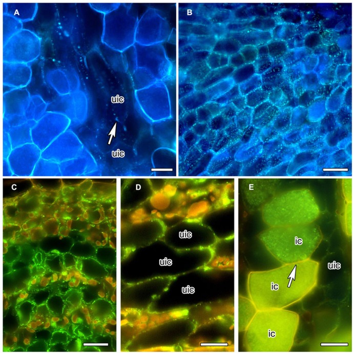
FIGURE 2.
Anilin Blue staining of callose (A,B) in longitudinal vibratome sections of living C. glauca nodules embedded in agarose (Brundrett et al., 1988), and immunolocalization of callose synthase (C–E) in cross sections of fixed nodules embedded in Steedman’s wax (for the method, see Zdyb et al., 2011; for the antibody, see Cai et al., 2011). Micrographs were taken under fluorescent light. The lignified walls of infected cells show in white under UV light (A,B) and in yellow under blue light (C–E) . (A,B) Punctate Anilin Blue-staining of callose (white fluorescence) is found in the walls connecting uninfected cells (uic; white arrow), but no labeling is found in infected cells [recognizable by their fluorescent cell walls; labeling should be visible because the fluorescence of Anilin Blue, as obvious in (B) which shows a younger area of the cortex before the onset of infection, is more yellowish then the fluorescence of the walls of infected cells]. (C) Callose synthase is detected in the plasma membranes of uninfected cortical cells in the youngest part of the cortex, close to the apical meristem. In the older part of the cortex – (D) shows the outer cortex in the zone of nitrogen fixation – the labeling is less dense. (E) No labeling was seen in the plasma membranes of infected cells (ic; arrow). Size bars denote 20 μm in (A–D) and 25 μm in (E) .

