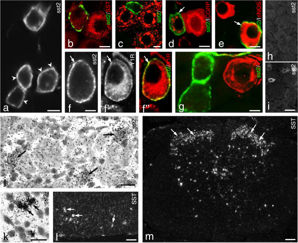
Figure 1.
sst2A-LI in mouse DRGs. (a) Several sst2A+ neurons are seen, and receptor protein is mainly located along the somatic plasmalemma (arrowheads). (b-e, f”,g) Color images show merged micrographs after double-staining (f-f” show the same section). (d-f”) Arrows indicate the coexistence of sst2A with CGRP (d), nNOS (e) and Y1R (f-f”), respectively. (b, c, g) sst2A-LI cannot be detected in SST+(b), IB4+(c) or Y2R+(g) neurons. CGRP-and nNOS-LIs are mainly seen in the perinuclear region (d, e), while Y1R-LI is found both in the plasmalemma and in the perinuclear region (f’, f”). (h, i) Lack of sst2A-LI in DRGs of sst2-KO mouse (h) but presence in WT mouse (i). (j, k) Expression of sst2 mRNA in DRG neurons (j, arrows) and spinal dorsal horn neurons (k, arrows). (l, m) Expression of SST mRNA in DRG neurons (l, arrows) and spinal dorsal horn (m, arrows). Scale bars indicate 10 μm (a-g, k), 20 μm (j), 50 μm (i, l) and 100 μm (m).

