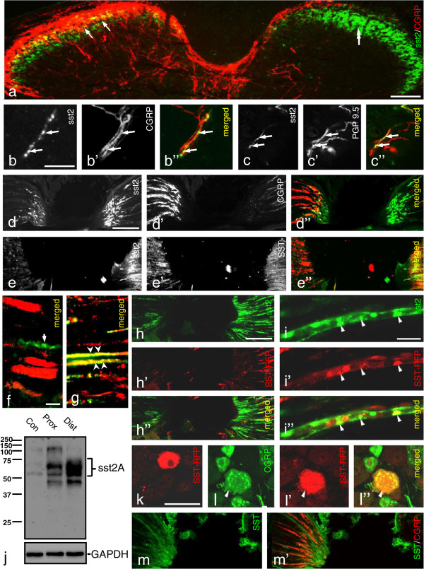
Figure 5.
Axonal transport of sst2A-LI. (a) Double-staining shows strong expression of sst2A-LI in the superficial dorsal horn, partially overlapping with the CGRP-LI (arrows in a). Fourteen days after unilateral dorsal rhizotomy, there is a dramatic reduction of CGRP-LI ipsilaterally (double-arrow in a) but not contralaterally (arrows in a). sst2A-LI seems unaffected. (b-c”) Skin of control mouse hindpaw. A few peripheral terminals of sciatic nerve contain sst2A-LI that co-localizes with CGRP (arrows in b-b”, same section) or PGP9.5 (arrows in c-c”, same section). (d-d”) A 10-hr ligation causes a modest accumulation of sst2A-LI on the proximal side, but a much stronger one distally (right), versus the mainly proximal CGRP pile-up (left) (cf. d’ with d; d”). (e-e”) SST-LI is present on both sides of the lesion (e’), and highly coexpressed with sst2A on the distal, but not on the proximal side (right) (cf. e’ with e; e”). Double-staining for sst2A and SST indicates possible coexistence on the distal (g; arrowheads) but not proximal side (f; an arrow points to an sst2A+ fiber lacking SST-LI). (j) Western blot showing higher sst2A protein levels on the distal than on the proximal side 10 hrs after ligation. (h-h”) Accumulation of sst2A and SST-RFP on the distal side after SST-RFP injection into the hind paw. (i-i”) The SST-RFP conjugate in distal axons is co-localized with sst2A (arrowheads). (k-l”) A few SST-RFP+ neurons are seen in ipsilateral DRGs 3 days after injection (k), most being CGRP+(l-l”). (m, m’) In sst2 KO mouse SST-LI only accumulates on the proximal side (contrasting the WT mouse, e’). CGRP-LI accumulates on the proximal side as see in WT mice (m’). Scale bars indicate 10 μm (f), 20 μm (i), 50 μm (k), 100 μm (a, b, d) and 200 μm (h).

