
Figure 7.
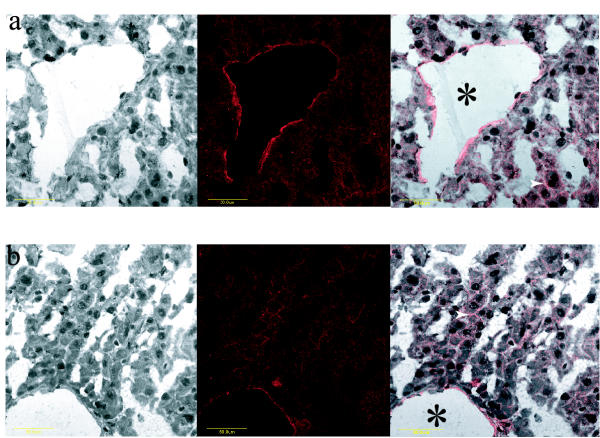
Immunohistochemical evaluation of mouse Abcg5/sterolin-1 expression in liver. Wild-type liver incubated with UTSW anti-Abcg5/sterolin-1 resulted in apical expression (panel a). Merged image clearly shows the apical distribution. Likewise Abcg8-/- liver incubated with the same UTSW anti-Abcg5/sterolin-1 antibody resulted in a similar apical expression pattern relative to the wild type (panel b). White arrows indicate bile canaliculi and asterisk indicates a bile duct. The yellow bar represents 50 μm.