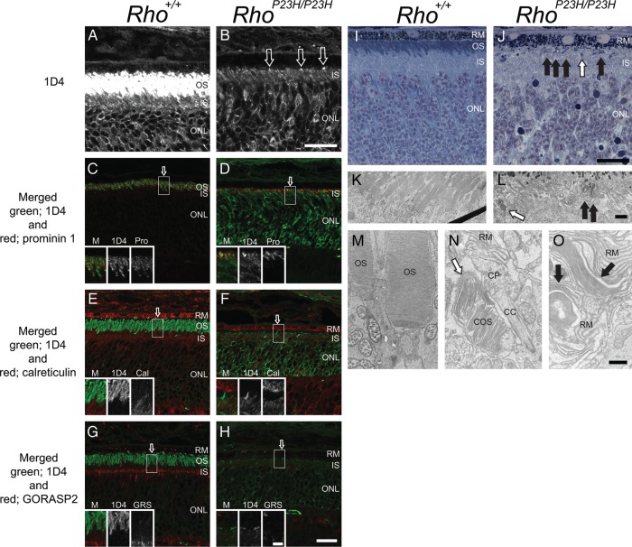
Figure 1.
Immunohistochemistry, light microscopy and TEM of Rho+/+ and RhoP23H/P23H mouse retinas suggest elongated disc formation in the RhoP23H/P23H mouse retina. Rho+/+ and RhoP23H/P23H mouse retinas at PND14 were analyzed with immunohistochemistry, light microscopy and TEM. Confocal microscopy conditions used to visualize the 1D4 signal in RhoP23H/P23H retinas (B) resulted in a saturated ROS signal in Rho+/+retinas (A) with staining also observed in the IS and ONL. 1D4 staining of RhoP23H/P23H retinas revealed concentrated needle or dot-shaped structures at the top of the IS (B, arrows) and within the IS layer (B). In Rho+/+ retinas, opsin and prominin 1 co-localized (C, yellow). In RhoP23H/P23H retinas, the prominin 1 signal co-localized with the concentrated needle-shaped opsin staining at the top of the IS (D, yellow). In Rho+/+ mouse retinas, opsin (E, green) did not co-localize with calreticulin in the IS or RPE (E, red). In RhoP23H/P23H mouse retinas, the concentrated needle-shaped opsin structures at the top of IS (F, green) did not co-localize with calreticulin (F, red). In contrast, the needle-shaped structures within the IS layer did co-localize with calreticulin (F, yellow). Opsin (green) did not co-localize with the Golgi reassembly stacking protein 2 (GORASP2) (red) in either Rho+/+ (G) or RhoP23H/P23H (H) mouse retinas. Higher magnification images of ROS regions indicated by arrows in (C)–(H) are shown in the insets. Light microscopy images of toluidine blue stained plastic sections are shown in (I) and (J). In Rho+/+ mouse retinas, OS appeared as blue or purple (I, OS). In RhoP23H/P23H mouse retinas, in addition to purple colored short OS (J, white arrow), purple colored abnormal OS structures were detected between the IS and RPE (J, black arrows). Thin sections from the same retina shown in (I) and (J) were analyzed by TEM (K and M, L, N and O for each). In Rho+/+ mouse retinas, stacks of discs (K and M) were observed. In RhoP23H/P23H mouse retina, COS (L and N, white arrows), a CP (N) and packs of elongated discs (L and O, black arrows) are shown. OS, photoreceptor cell outer segment; IS, photoreceptor cell inner segment; ONL, outer nuclear layer; M, merged image; Pro, prominin 1; Cal, calreticulin; GRS, GORASP2; RM, retinal pigment epithelium (microvilli); COS, cone photoreceptor cell outer segment; CP, ciliary protrusion; CC, connecting cilium/cilia. Scale bar: 20 µm (A–J); 5 µm (C–H, insets); 2 µm (K and L); 500 nm (M–O).

