Figure 3.
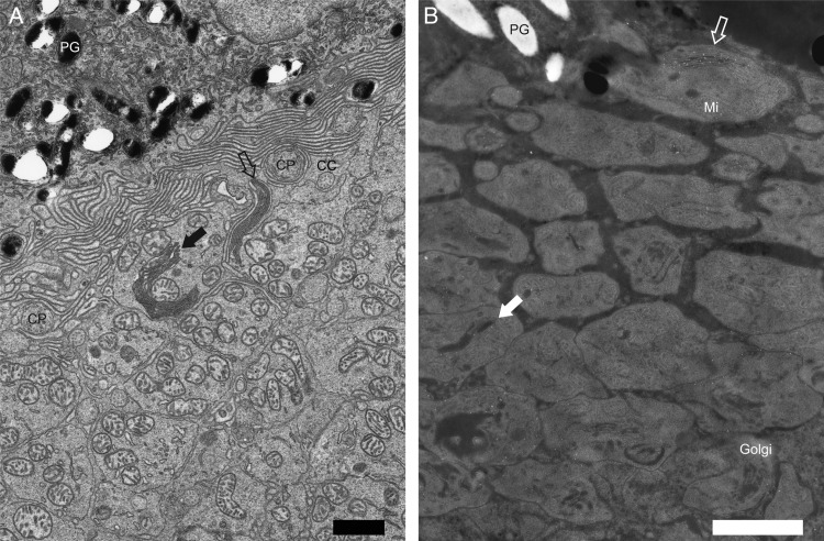
Low magnification images of Figure 2C and F. Low magnification images of Figure 2C (A) and Figure 2F (B). RhoP23H/P23H mice featured elongated discs (A and B,  ) or disc-like structures inside the IS (A and B,
  and solid white
  ). In contrast to elongated discs (A,
  ), disc-like structures inside the IS were located near the center of the IS (A,
  ). Opsin was predominantly located at the elongated discs (B,
  ), which were adjacent to the IS and retinal pigment epithelium. Less intense opsin staining was also detected at disc-like structures inside the IS (B, solid white
  ). Black spaces within IS disc-like structures (B, solid white
  ) likely correspond to empty space, an artifact of immuno-EM sample preparation. In the Rho+/+ mouse immuno-EM samples, a similar artifact was often observed between discs (Fig. 2D). PG, pigment granule of the RPE; Golgi, Golgi apparatus. Other abbreviations are the same as in Figure 2. Scale bar: 1 µm.

