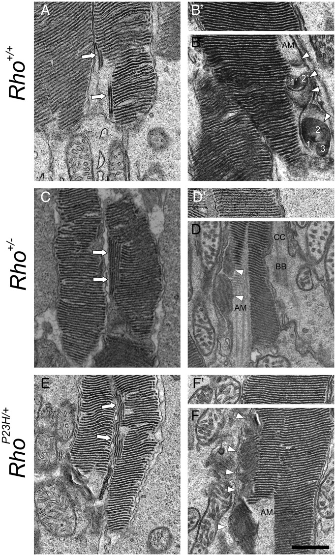
Figure 6.
Retinal TEM images of sagittal sections of PND14 mouse photoreceptor cell outer segments demonstrate sagittally oriented small discs adjoining normal stacks of discs. Sagittally oriented small discs were detected as double membranes ( ) or discs (Δ) in photoreceptor sagittal sections (A–F). Those discs were next to stacks of normally oriented discs (A, C and D), the AM and stocks of normally oriented discs (B, D and F). (B), (D) and (F) show transverse sectional views of sagittally oriented small discs located close to proximal ends of outer segments. Note that two sagittally oriented small discs in (B) exhibit a trace of two (1′, 2′) or three smaller discs (1,2,3) with (B′), (D′) and (F′) showing distal parts of same outer segment. At the distal ROS, these discs are enlarged but normally oriented. CC, connecting cilium; AM, axonemal microtube; BB, basal body. Scale bar: 500 nm.

