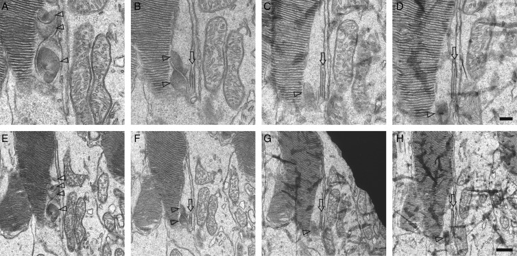
Figure 7.
Retinal TEM images of serial sections from Rho+/+ mouse retinas reveal a series of sagittally oriented discs. TEM images are shown of four serial sections prepared from Rho+/+ mice at PND14 (A–D). These covered a thickness of ∼320 nm of photoreceptor cells or ∼80 nm for each section. (E–H) Lower magnification images of (A)–(D). Note that small sagittally aligned disc(s) are shown in four of four serial sections at the proximal part of the same photoreceptor cell (Δ). Also, sagittally oriented three double membranes ( ) are present in three of the four serial sections. Scale bar: 200 nm (A–D) and 500 nm (E–H).

