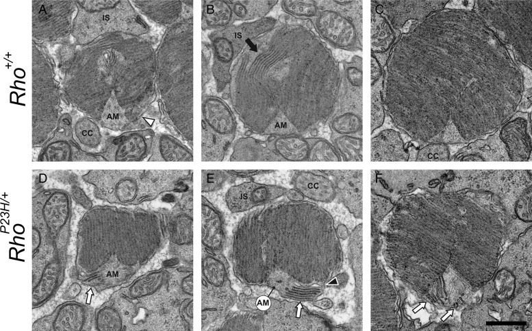
Figure 9.
TEM images of transverse sections of PND63 mouse photoreceptor cell outer segments reveal sagittally oriented discs near the axonemal microtube (AM) in RhoP23H/+ mice but not in Rho+/+ mice. At PND63, mature photoreceptor cells continuously synthesize new discs at the proximal outer segment where the opposite side of the AM is surrounded by the distal inner segment (A, IS). In Rho+/+ mice at PND63, sagittally aligned discs were not seen around the AM (A–C), but transversely aligned small discs were detected instead (A, Δ). Also, a different type of sagittally aligned disc was located at the side opposite the AM (B,  ). Routinely observed, this type of disc did not display a rim and both ends of it were connected to transversely aligned discs, indicating that these sagittally aligned discs represented partially folded transversely aligned discs at the proximal part of the ROS. In RhoP23H/+ mice, sagittally aligned discs were still seen around the AM (D–F,
  ). The open disc indicated by a black arrowhead in (E) was probably an artifact caused by sample preparation. Similar types of open discs were also noted in the ROS of Rho+/+ mouse retinas (data not shown). Abbreviations are as in Figure 6. Scale bar: 500 nm.

