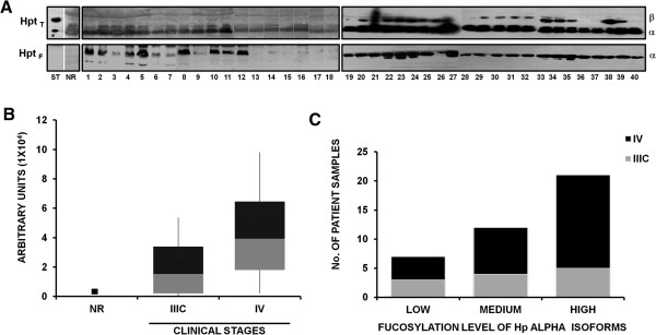
Figure 3.

Fucosylation profile of haptoglobin. A). Haptoglobin standard (ST), an ascitic fluid sample non-related with cancer (NR), and 40 ascitic fluid samples from ovarian cancer patients were analyzed for fucosylation of haptoglobin alpha. Protein samples (50 μg) from ascitic fluid free of abundant proteins were processed through a 12.5% SDS-PAGE and transferred to a NCP to incubate with biotinylated-Aleuria aurantia lectin (1:1000), specific for fucose residues; variable levels of fucosylation were detected by a colorimetric system using Alkaline Phosphatase-conjugated Streptavidin (1:2000) (lower left panel) or with Horseradish Peroxidase-conjugated Streptavidin (1:1000) (lower right panel). As loading control, the same samples were incubated with an anti-Hp antibody that recognizes the β, α1 and α2 isoforms of Hp to detect total Hp, and a secondary antibody conjugated with Horseradish Peroxidase (1:1000) (upper right panel) or Alkaline Phosphatase-conjugated antibody (1:1000) (upper left panel). B). Graph showing densitometric values of samples’ fucosylation in arbitrary units, comparing clinical stages IIIC and IV vs. a non-cancer related sample. C). Fucosylation levels (low, medium or high) of samples, according to clinical stages.
