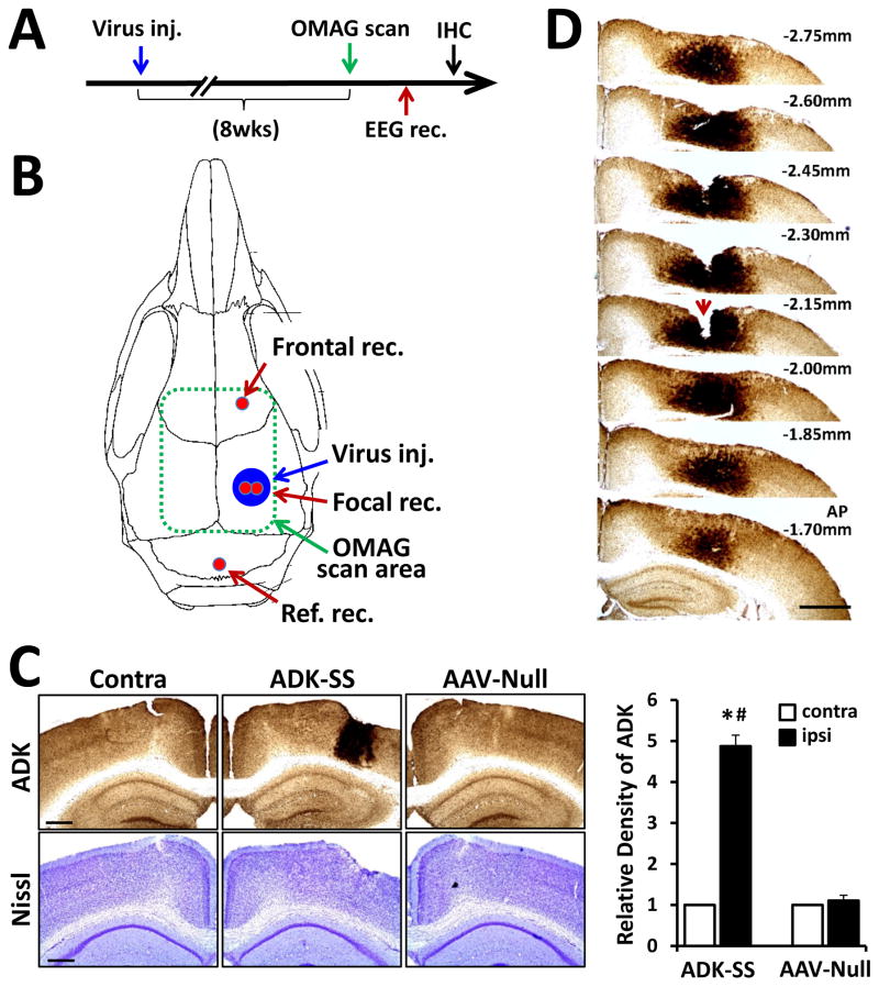
Fig. 1.
Overexpression of ADK in the neocortex of mice induced by unilateral injection of ADK sense (ADK-SS) virus. A: Schematic illustration of the experimental paradigm. Mice were first subjected to unilateral intracortical virus injection, followed after 8 weeks by an OMAG scan and cortical EEG recordings. After completion of the EEG, animals were sacrificed for immunohistochemistry analysis. B: Schematic illustration of the virus transduced area (Virus inj.) locations of the EEG recording electrodes (red): in frontal cortex (Frontal rec.), focal area of ADK overexpression (Focal rec.), and above the cerebellum (Ref. rec.). The area scanned by OMAG is indicated by a dashed green line. C: Representative ADK immunoperoxidase (upper panel) and Nissl (lower panel) staining of brain slices showing the contralateral (Contra) or ipsilateral injected (ADK-SS) hemisphere of an ADK-SS recipient and the virus-injected hemisphere of an AAV-Null injected control animal (AAV-Null). The bar graph demonstrates relative densities of ADK immunoreactivity of DAB-stained brain sections of ADK-SS or AAV-Null virus-injected mice (two sections per animal, n=5 animals per group), scale bar = 500 μm. D: Representative ADK staining of consecutive brain slices from an ADK-SS virus-injected animal showing the three dimensional extent of ADK overexpression. Notch (arrow) on −2.15 mm slice indicates the locus of the electrode implantation, scale bar = 1 mm. Data are displayed as mean ± SEM. # P<0.01 ADK-SS vs AAV-Null group; * P<0.01 ipsilateral vs contralateral side. Abbreviations: Virus inj. = virus injection area; Ref. rec= reference electrode recording; Frontal rec = frontal electrode recording; Focal rec. = electrode recording; contra = contralateral; ipsi = ipsilateral; IHC = immunohistochemistry

