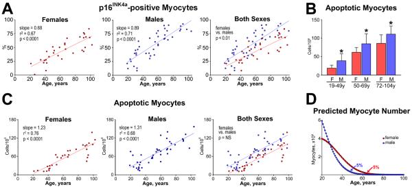
Fig. 3.

(A) The rate of increase in the fraction of p16INK4a-positive myocytes with age is lower in the female than in the male left ventricle (LV). (B) The fraction of apoptotic myocytes in the LV is lower in women than men. F indicates female; M, male. *P<0.05 vs. female. (C) The rate of increase of apoptotic myocytes with age is similar in the female and male LV. (D) At 20 years of age, the female (red) and male (blue) LV contains 4 and 6×10⁹ cardiomyocytes, respectively. Curves are derived by combining the yearly levels of apoptosis shown in panel C and the prediction that cell death lasts 4 hours. In the absence of cell regeneration, 5% of LV myocytes (200×106 in the female LV and 300×106 in the male LV) would be left at 63 and 48 years of age in the female and male LV, respectively. Figure adapted from reference [23].
