
Fig. 4.
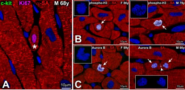
(A) Amplifying myocytes (asterisk) are small cycling cells, positive for Ki67 (magenta) and lack c-kit. (B, C) Small amplifying myocytes are positive for phospho-H3 (B, arrow) (B) and aurora B kinase (C, arrow). In the right panel in C, aurora B kinase is located in the 2 sets of telophase chromosomes and at the cleavage furrow of the dividing myocyte (arrowhead). Insets in B and C illustrate the organization of chromosomes in the dividing cells. Figure adapted from reference [23].