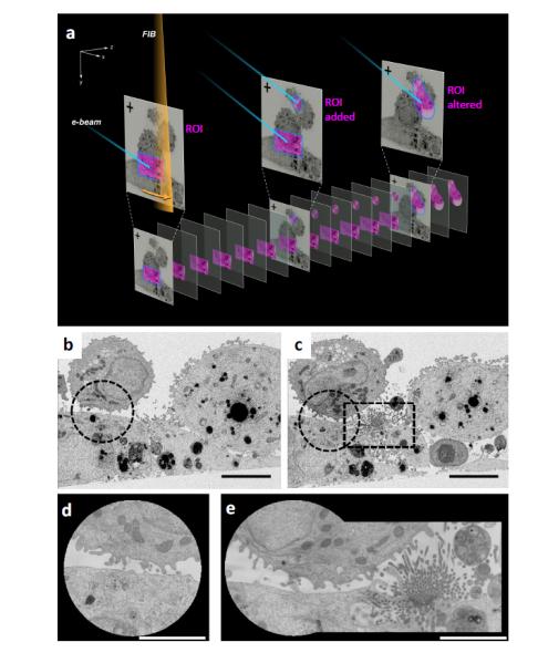
Figure 1. Targeted 3D FIB-SEM by keyframe imaging.

(a) FIB milling (orange beam) and SEM imaging (blue beam) of resin embedded cells generates two image classes. Lower resolution keyframes of the entire field of view (top) are collected at sparse z intervals, and higher resolution images of ROIs (pink) are collected more frequently. The shape and number of ROIs can be altered during a run. (b,c) Keyframes of mammalian cell-cell contacts reveal a new area of interest midway through the experiment, ROI sub-areas indicated with dotted lines. (d) High resolution ROI image of initial synapse, (e) ROI image altered to include new area of interest.
