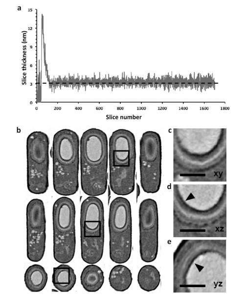
Figure 2. High resolution in 3D by controlled FIB milling.

(a) Plot of slice thickness over time during an overnight run of resin-embedded B.subtilis bacteria; the user defined thickness was 3 nm (dotted line). The calculated slice thickness measurements during autofocus and stigmation procedures were excluded. (b) Resampled slices through a representative B.subtilis bacterium, with orthogonal planes oriented to the principal axes of the cylindrical cell (xy: top; xz: middle and yz: bottom). Quality of data is comparable across all planes (c-e) Zoomed in images of the same region of the cell shown as slices in xy, xz and yz planes corresponding to the boxed region of the images in (b), revealing at least five clear concentric spore coat layers around the dark spore core. Small 20-30 nm electron dense features coating the core (arrowheads) can be resolved. Scale bar 400 nm.
