Fig 2.
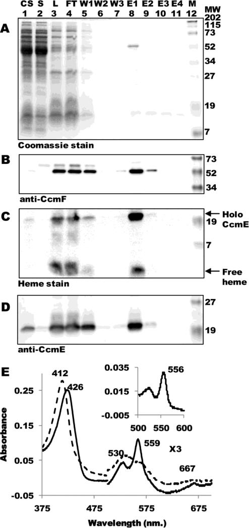
The CcmF-holoCcmE complex. (A) Coomassie blue staining of purified CcmF:His6 showing 54-kDa CcmF. (B) Anti-CcmF immunoblot of purified CcmF:His6 showing 54-kDa CcmF. (C) Heme staining of purified CcmF showing free heme (CcmF b-heme) and co-purified 20-kDa holoCcmE. (D) Anti-CcmE immunoblot showing co-purified 20-kDa CcmE. For (A)-(D), abbreviations are CS, crude sonicate; S, soluble fraction; L, load (DDM-solubilized membranes); FT, flow through; W1, wash 1; W2, wash 2; W3, wash 3; E1, elution 1; E2, elution 2; E3, elution 3; E4, elution 4; M, molecular weight standards. (E) UV-Vis absorption spectra of CcmF-holoCcmE complex as purified (dotted line) or reduced with sodium dithionite (solid line). The region from 500-700 nm has been multiplied by a factor of 3. (Inset) Sodium dithionite-reduced pyridine hemochrome spectrum of purified CcmF-holoCcmE complex from 500-600 nm. Absorption maxima are indicated.

