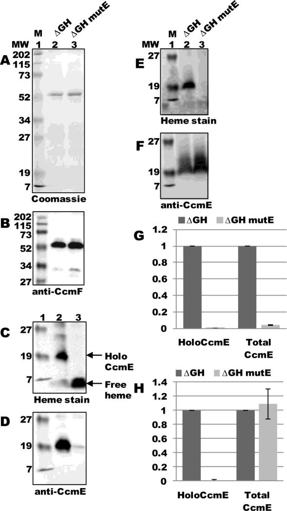
Fig 5.
ApoCcmE does not co-purify with CcmF. (A) Coomassie blue staining of TALON-purified proteins from ΔGH and ΔGH mutE (CcmE His130Ala) backgrounds showing purified 54-kDa CcmF. (B) Anti-CcmF immunoblot of purified CcmF proteins showing 54-kDa CcmF. (C) Heme staining of purified CcmF proteins showing free heme (CcmF b-heme) and co-purified 20-kDa holoCcmE. (D) Anti-CcmE immunoblot of purified CcmF proteins showing co-purified 20-kDa CcmE. For (A)-(D), 5 ug purified protein was analyzed. (E) Heme staining of DDM-solubilized membrane fractions from ΔGH and ΔGH mutE backgrounds showing 20 kDa holoCcmE. (F) Anti-CcmE immunoblot of DDM-solubilized membrane fractions showing 20 kDa CcmE. For (E) and (F), 70 ug total protein was analyzed. (G) Quantification of the results of heme staining (holoCcmE) and anti-CcmE immunoreactivity (total CcmE) from purified fractions from three independent experiments. (H) Quantification of the results of heme staining and anti-CcmE immunoreactivity from DDM-solubilized membrane fractions from three independent experiments. For (G) and (H), percent holoCcmE and total CcmE is relative to ΔGH, which has been set at 100%. Error bars denote SD.

