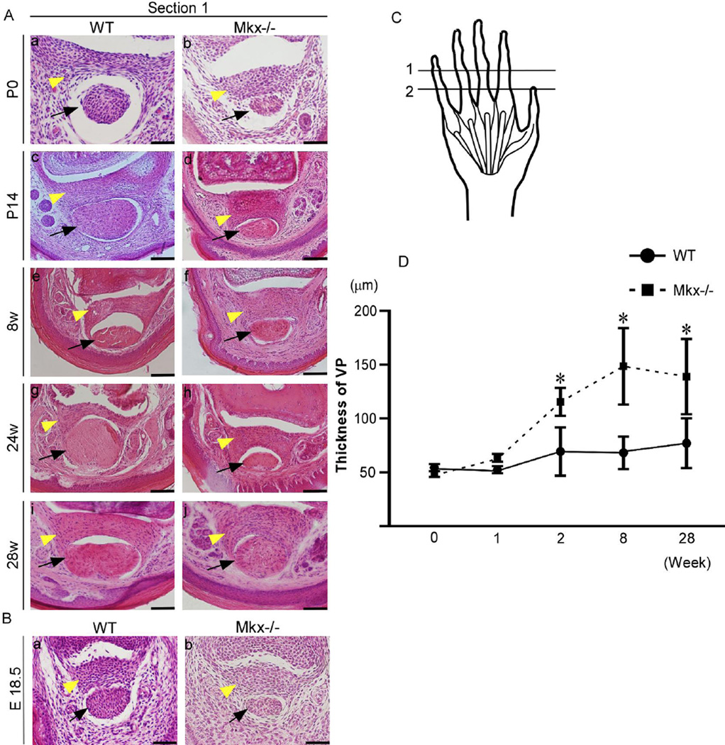
Figure 5. Tendon mass decreases in Mkx-null mice since E18.5. Volar plates (VPs) are normal in embryonic Mkx-null mice and become thick in Mkx-null mice after P14.
(A) Hematoxylin and eosin (H&E) staining of the flexor digitorium profundus (FDP) tendon and VP in wild-type (a, c, e, g, and i) and Mkx-null mice (b, d, f, h, and j), in the time range from P0 to 28 weeks. Scale bars are 50 µm in a and b and 100 µm in c–j. (B) H&E staining of the FDP tendon and VP in E18.5 wild-type (a) and Mkx-null mice (b). Scale bar, 50 µm. Yellow arrowheads, VPs. Black arrows, FDP tendons. (C) Section levels are marked by black lines: (1) proximal interphalangeal (PIP) joint, (2) middle of the proximal phalanx (level of the A2 pulley). The sections were taken at level 1. (D) Comparison of the thickness of VPs between Mkx-null mice and wild-type littermates. *P < 0.05, n = 5.

