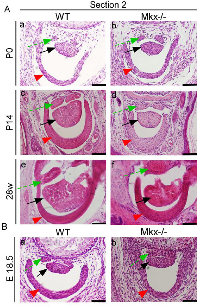
Figure 6. Pulleys are normal in embryonic Mkx-null mice and become thick in Mkx-null mice after P14.
(A) Hematoxylin and eosin (H&E) staining of the flexor digitorium profundus (FDP) tendon, flexor digitorum sublimis (FDS) tendon, and pulleys in wild-type (a, c, and e) and Mkx-null mice (b, d, and f) in the time range from P0 to 28 weeks. Scale bars are 50 µm in a and b and 100 µm in c–f. (B) H&E staining of the FDP tendon, FDS tendon, and pulley in E18.5 wild-type (a) and Mkx-null mice (b). Scale bar, 50 µm. Green dashed arrows, FDS tendons. Red arrowheads, pulleys. Black arrows, FDP tendons. The sections were taken at level 2 (for details, see the caption of Fig. 5-C).

