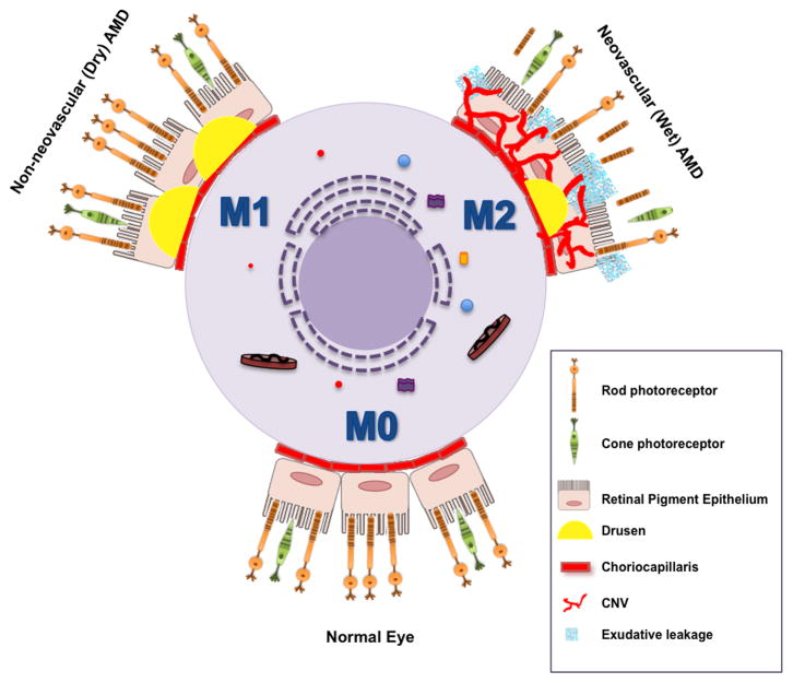
Figure 2. The central role of macrophage-mediated immunity in disease pathogenesis of AMD.
Under baseline conditions, non-polarized macrophages (M0) traffic through the choriocapillaris underneath the retina sampling the tissue microenvironment. Aging and complex genetic factors impair macrophage function including reverse cholesterol transport (RCT) and lead to the deposition of lipid rich drusen underneath the retina and RPE in non-neovascular (dry) AMD. In dry AMD, dysfunctional classically activated macrophages (M1) can also induce inflammation driven cell death that leads to advanced stages of dry AMD characterized by loss of RPE cells, called geographic atrophy (GA) and subsequent loss of rod and cone photoreceptor neurons (PR). In the more aggressive form of disease, impaired macrophage RCT in the drusen rich sub-retinal micromilieu polarizes these cells to an alternatively activated (M2) phenotype. Alternatively activated macrophages are disease promoting and pro-angiogenic and facilitate the development of CNV. The sine qua non of wet AMD is the development of CNV, characterized by the growth of new blood vessels from the choroid into the sub-retinal space. These neovascular complexes are leaky and lead to exudation and hemorrhage with disruption of the retinal architecture, interference with the central visual axis, loss of PR and ultimately, irreversible vision loss.

