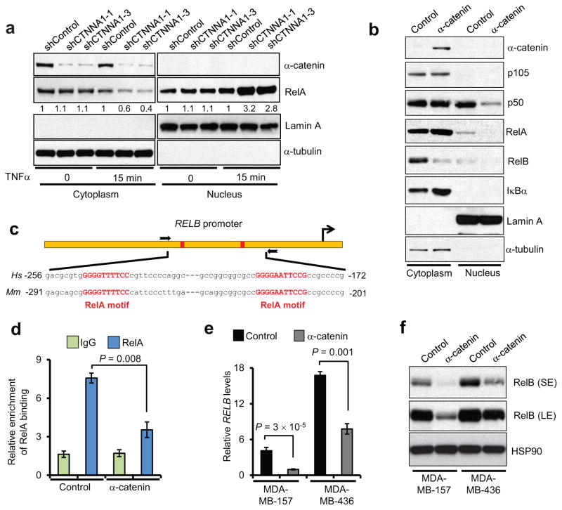
Figure 4. α-catenin inhibits RelA-p50 nuclear localization and downregulates RelB.
(a) Immunoblotting of α-catenin and RelA in cytoplasmic and nuclear fractions of BT549 cells transduced with two independent α-catenin shRNAs, with or without TNFα treatment.
(b) Immunoblotting of α-catenin, p105, p50, RelA, RelB and IκBα in cytoplasmic and nuclear fractions of α-catenin-transduced MDA-MB-157 cells. α-tubulin and Lamin A were used as cytoplasmic and nuclear markers, respectively, in (a) and (b).
(c) Schematic representation of the RELB promoter containing two RelA binding sites (red rectangles). The two boxed arrows indicate the primers used for ChIP-qPCR. Hs: Homo sapiens, Mm: Mus musculus.
(d) ChIP-qPCR analysis of RelA binding to the RELB promoter in α-catenin-transduced MDA-MB-157 cells. qPCR was performed with primers specific to the RelA binding motifs. Data were normalized to the input. n = 3 samples per group.
(e) qPCR of RELB in α-catenin-transduced MDA-MB-157 and MDA-MB-436 cells. n = 3 samples per group.
(f) Immunoblotting of RelB and HSP90 in α-catenin-transduced MDA-MB-157 and MDA-MB-436 cells. SE: short exposure; LE: long exposure.
Data in (d) and (e) are the mean of biological replicates from a representative experiment,, and error bars indicate s.e.m. Statistical significance was determined by a two-tailed, unpaired Student’s t-test. The experiments were repeated three times. The source data can be found in Supplementary Table 4. Uncropped images of blots are shown in Supplementary Fig. 7.

