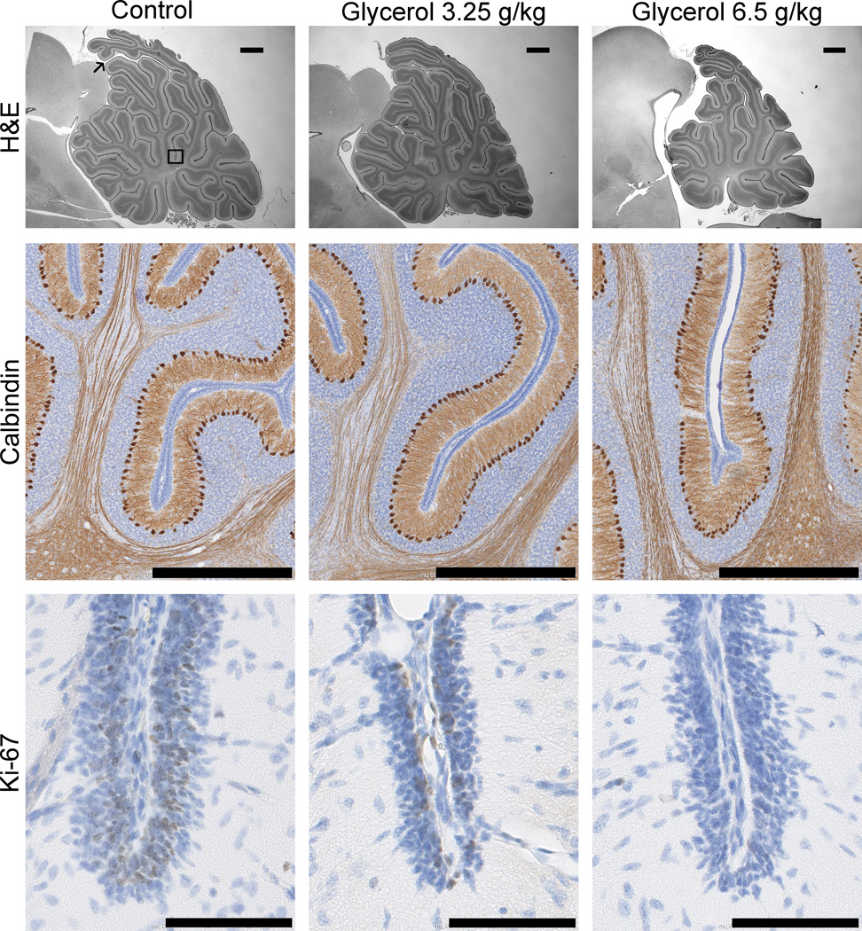
Figure 3.
Photomicrographs showing the dose-dependent effects of systemic glycerol treatment on cerebellar development. Brain sections from control (left column) and glycerol-treated (right columns) rabbit kits were stained for H&E (top row, scale bar 1 mm), calbindin-D28K (middle row, scale bar 800 µm) and Ki-67 (bottom row, scale bar 90 µm). Measurements were made along the primary fissure of the vermis, as denoted in the upper left panel by the thin black arrow marking the start, and the black box marking the end of the region of interest. Cerebellar growth was compromised because the density of calbindin-immunopositive Purkinje cells was increased, and the proportion of Ki-67-immunopositive proliferating external granular layer cells (thick black arrows) was decreased in glycerol-treated kits. Morphological measurements using H&E-stained images indicated that the foliation is less complex, and the thickness of the external granular layer is decreased at the base of the primary fissure in glycerol-treated kits.

