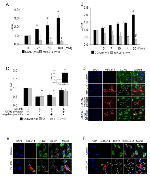
Fig. 2. MiR-214-dependency of CCN2 production in mouse HSC.
(A) RT-PCR of CCN2 mRNA or miR-214, normalized to GAPDH mRNA, after Day 3 primary mouse HSC were incubated in 1% serum for 24 hrs prior to 48-hr treatment with 0–100mM ethanol (n=5 independent experiments performed in triplicate, *P <0.001 vs. non treatment, +P <0.01 vs. non treatment). (B) CCN2 mRNA or miR-214 expression, assessed by RT-PCR and normalized to GAPDH mRNA, over 20 days of primary culture of HSC isolated from normal mouse liver (n=4 independent experiments performed in triplicate, *P <0.001 vs. Day 1 CCN2 expression, #P <0.001 vs. Day 1 miR-214 expression). (C) RT-PCR of CCN2 or collagen α1(I) mRNA or miR-214 (inset) relative to GAPDH mRNA in RFP-positive HSC collected by cell sorting or (D) detection of CCN2 protein by indirect immunofluorescence (green) or RFP by direct fluorescence (red) in P6 HSC transfected for 24 hrs with pLemiR-214 either alone or with a co-transfected CCN2 3′-UTR protector. A negative protector was used as a control (n=4 independent experiments performed in triplicate; *P<0.001 vs control). RFP fluorescence (red) and indirect immunofluorescence for CCN2 (green) and (E) αSMA (white) or (F) collagen α1 (white) in P6 control HSC or after transfection for 24 hrs with pLemiR-214. In (D–F), blue is DAPI nuclear stain. Scale bar: 20μm.

