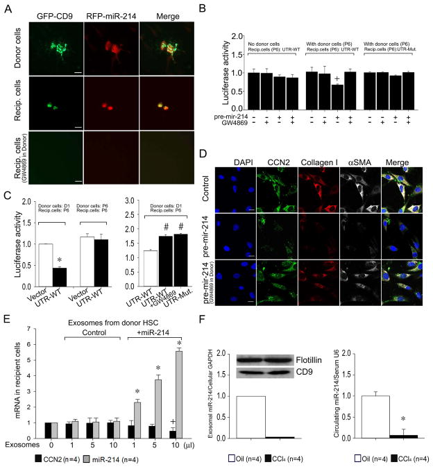
Fig. 5. HSC-HSC intercellular signaling via exosomal transport of miR-214.
(A) HSC donor and recipient co-culture experiments were performed as shown in Supporting Fig. S4. Donor P6 mouse HSC were co-transfected with plasmids for GFP-CD9 (green) and RFP-miR-214 (pLemiR-214; red) and cultured with or without GW4869. Recipient cells were non-transfected P6 mouse HSC. Representative immunofluorescence images of the donor and recipient cells are shown (n=3 independent experiments.) Scale bar: 20μm. (B) Recipient P6 mouse HSC transfected with Fire-Ctx plasmids containing either wild-type or mutant CCN2 3′-UTR (Fig. 3A,B, Supporting Figs. S1A, S4B) were co-incubated with control or pre-mir-214-transfected donor P6 HSC in the presence or absence of GW4869. In control experiments, some recipient HSC were exposed to the donor side of the micro-wells which lacked donor HSC but contained pre-mir-214 in the culture medium at the same concentration (100nM) as used for donor HSC transfection. The activity of firefly luciferase in lysates of recipient cells, normalized to that of Renilla luciferase, is shown 24 hrs after the central divider had been removed from the micro-wells. (n=4 independent experiments performed in triplicate, +P <0.05 vs. control group). (C) HSC co-culture experiments were set up and evaluated as in (B) except donor HSC had been freshly isolated from normal mouse livers and placed in culture for 1 day prior to treatment with GW4869 (n=4 independent experiments performed in triplicate, *P <0.001 vs. vector, # P <0.001 vs. UTR-WT). (D) Co-cultures were established for 12 hrs between control or pre-mir-214-transfected P6 donor HSC (± GW4869) and P6 recipient cells (Supporting Fig. S4B), the latter of which were then evaluated for immunocytochemical detection of CCN2 (green), collagen I (red) or αSMA (white). Blue depicts DAPI nuclear stain. Scale bar: 20μm. (E) Expression of CCN2 or miR-214 in P6 HSC after addition for 24hrs of exosomes isolated from 48-hr conditioned medium of control P6 HSC or P6 HSC transfected with pre-mir-214. (F) MiR 214 in exosomes from 24-hr conditioned medium of HSC isolated from Swiss Webster mice receiving two injections of oil or CCl4 (Western blots show detection of flotillin or CD9 in exomosal extracts) (left) or serum from Swiss Webster mice that were administered oil or CCl4 for 5 weeks (right) (*P < 0.001 vs oil group).

