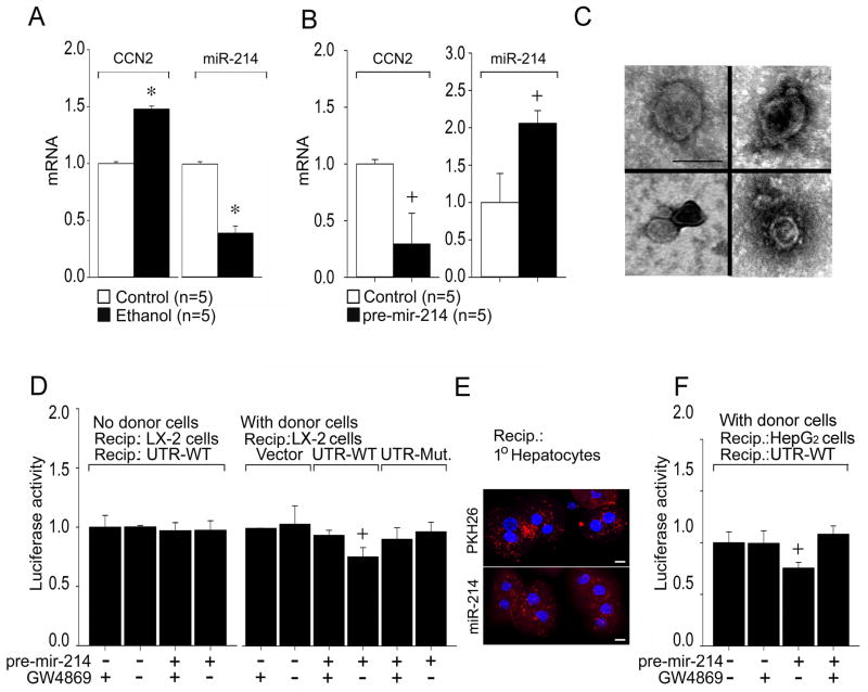
Fig. 6. Functional characterization of miR-214 produced by human HSC.
(A) Reciprocal expression of CCN2 and miR-214 after serum-starved LX-2 HSC were treated for 48 hrs with 25mM ethanol. RNA was subjected to RT-PCR and expression of CCN2 or miR-214 was normalized to that of GAPDH. (n=5 independent experiments performed in triplicate, * P<0.001 vs non treatment). (B) Down-regulation of CCN2 expression in LX-2 cells after transfection with pre-mir-214, determined as in (A). (n=5 independent experiments performed in triplicate, +P<0.05 vs Control). (C) TEM of exosomes purified from LX-2 cell conditioned medium. Scale bar: 100nm. (D) Donor LX-2 cells were transfected with pre-mir-214, in the presence or absence of GW4869. Some donor wells contained pre-mir-214 in the medium but no LX-2 cells. Recipients were LX-2 cells transfected with Fire-Ctx plasmids that were parental or contained wild-type or mutant CCN2 3′-UTRs (Supporting Fig. S4B). Firefly luciferase activity was measured in cell lysates 24 hrs after co-culture and normalized to that of Renilla luciferase. (n=4 independent experiments performed in triplicate, +P <0.05 vs. control group). (E) Confocal microscopy showing uptake of exosomes by primary mouse hepatocytes after 4-hour incubation with P6 HSC-secreted exosomes pre-labeled with PKH26 (upper) or that were isolated from the medium of P6 HSC transfected with pLemiR-214 (lower); Scale bar: 10μm. (F) Co-cultures were established for 24 hrs between pre-mir-214-transfected donor LX-2 cells (± GW4869) and recipient HepG2 cells transfected with Fire-Ctx plasmid containing wild-type CCN2 3′-UTR (Supporting Fig. S4B). Luciferase was measured in cell lysates as in (D). (n=4 independent experiments performed in triplicate, +P <0.05 vs. control group).

