
Figure 1. Insulin stimulated GLUT4 glucose transporter translocation.
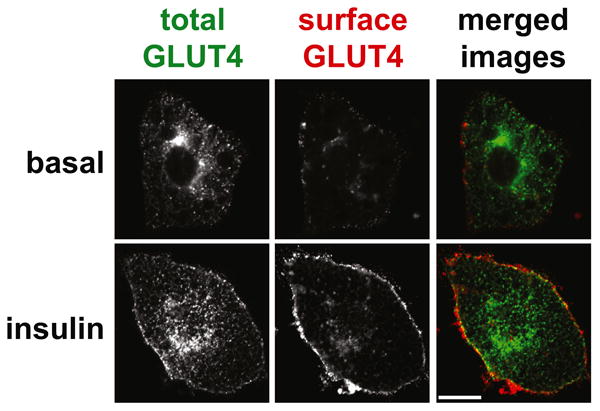
The images show cultured 3T3-L1 adipocytes that express a GLUT4 reporter protein, which contains a 7Myc epitope tag in its first extracellular loop as well as GFP fused at the C-terminus. Cells were serum starved, treated with or without insulin as indicated, then stained to detect the externalized 7Myc epitope tag. Images were acquired by confocal microscopy of GFP (total GLUT4, shown in green in the merged images) and Myc epitope (surface GLUT4, shown in red in the merged images). Scale bar, 10 μM. Reproduced from Yu, C., et al., J Biol Chem (2007) 282, 7710-7722.