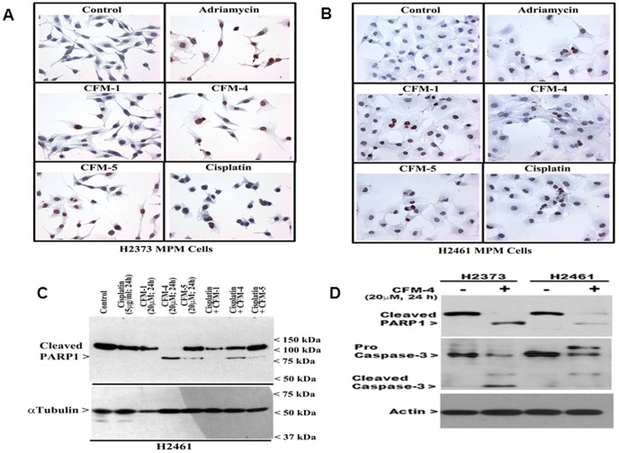
Figure 2. CFMs stimulate apoptosis in MPM cells.
(A, B) Indicated MPM cells were either untreated (Control), treated with 2.5 µg/ml Adriamycin, 5 µg/ml Cisplatin or 20 µM dose of respective CFMs for 24 h. Staining of the cells was performed using TUNEL assay as detailed in Methods. Dark brown staining represents fragmented cell nuclei. (C, D) MPM cells were either untreated (denoted as Control in panel C and – in panel D) or treated (denoted as+in panel D) with indicated agents for noted time and dose, and levels of cleaved PARP, pro- and cleaved (activated) caspase-3, and actin proteins were determined by Western blotting.

