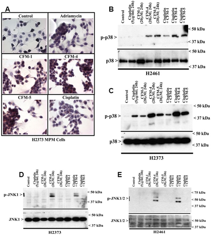
Figure 4. CFMs activate pro-apoptotic SAPKs in MPM cells.
(A, B) Indicated MPM cells were either untreated (Control), treated with Adriamycin, Cisplatin, or respective CFMs as in figure 2A. Staining of the cells was performed using anti-phospho-p38 antibody as detailed in Methods. Presence of p38 is indicated by intense brown staining in the nuclei and cytosol of the treated cells. MPM cells were either untreated (Control) or treated with indicated agents for noted time and dose, and levels of phosphorylated p38 (noted as p-p38), and total p38 proteins (B, C) or phosphorylated JNK (noted as p-JNK1/2), and total JNK proteins (D, E) were determined by Western blotting essentially as in figure 2.

