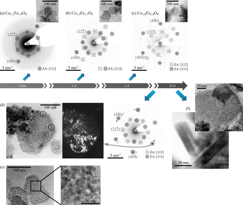
Figure 3.
Top: Change in the electron diffraction pattern of diamond shaped particles with time. The inverted electron diffraction pattern is given here. (a) t = 5 min, (b) t = 1 d, (c) t = 2 d. Bottom: HRTEM images of incomplete particles. (d) Dark field image of an incomplete diamond-shaped particle as well as the corresponding electron diffraction pattern of the disc. The highlighted areas in the dark field image correspond to the specific reflex of the electron diffraction pattern. The inset of (e) shows an enlarged area within the disc. This disc is composed of smaller subunits.(f) HRTEM image of a final, stoichiometric monocrystalline disc, obtained after 28 days. The inset shows that the final nanoparticles are not porous. Only the dominating reflexes are indexed for reasons of clarity.

