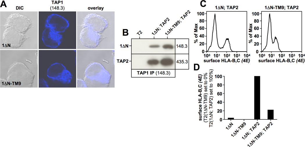
Figure 3. 1ΔN-TM9 localizes to the endoplasmic reticulum and retains function to a significant extent.
A) Immunofluorescence analysis of the indicated T2 transfectants using the TAP1-specific antibody 148.3. B) TAP1 was immunoprecipitated with antibody 148.3 from a digitonin lysate of the stable T2 transfectant expressing 1ΔN-TM9. Isolated proteins were analyzed by Western blotting using antibodies against TAP1 (148.3) and TAP2 (435.3). C, D) Flow cytometry analysis of surface-HLA-B,C expression using antibody 4E in the indicated T2 transfectants (C). Surface-HLA-B,C levels are shown as a bar diagram (1ΔN; TAP2 set to 100%) (D).

