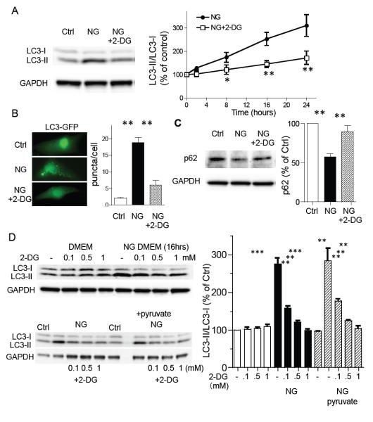
Figure 1. Glucose deprivation induced autophagy is inhibited by 2-deoxy-D-glucose (2-DG) in neonatal rat ventricular myocytes (NRVMs).
(A) Cardiomyocytes were cultured in DMEM or no-glucose (NG) DMEM in the presence or absence of 2-deoxy-D-glucose (2-DG; 0.5 mM) for 16 hrs and subjected to Western blotting for LC3 (left panels). The time-course of the LC3-II/LC3-I ratio induced by glucose deprivation, plus or minus 2-DG (0.5 mM), assessed by Western blotting (right panel; n=8-10). *P<0.05, **P<0.01 vs each time point after glucose deprivation. (B) To visualize formation of autophagy, NRVMs were infected with LC3-GFP adenovirus. After 24 hrs, cells were subjected to glucose deprivation (NG) in the presence or absence of 2-DG (0.5 mM) for 16 hrs. (C) Western blotting for p62. Cells were cultured in DMEM or no-glucose (NG) DMEM in the presence or absence of 2-deoxy-D-glucose (2-DG; 0.5 mM) for 16 hrs. **P<0.01 (n=6). (D) Cardiomyocytes were cultured in DMEM or NG-DMEM in the presence or absence of 2-DG (0.1, 0.5 and 1 mM). To energize mitochondria, 5 mM pyruvate was added (lower blots). **P<0.01, ***P<0.001 n=7. Data are mean ± SEM.

