
Figure 6. HK-II associates with TORC1 and this is increased by glucose deprivation.
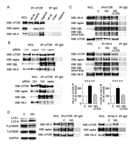
(A) Association of HK-II with mTOR is sensitive to the detergent. Cells were harvested in 0.5 % NP-40 lysis buffer, 0.3% Chaps lysis buffer or 0.02% digitonin lysis buffer, subjected to mTOR immunoprecipitation and Western blotting for HK-II and raptor. (B) Knockdown of raptor decreases the association of mTOR with HK-II but not with GβL (upper panels). HK-I does not associate with mTOR (lower panels). NRVMs transfected with control siRNA or raptor siRNA were subjected to glucose deprivation for 16 hrs and mTOR immunoprecipitation was carried out. (C) Glucose deprivation increases association between HK-II and TORC1 which is inhibited by 0.5 mM 2-DG. NRVMs were subjected to glucose deprivation ± 2-DG for 16 hrs and mTOR or HK-II were immunoprecipitated. Right panels show quantitative analysis of the association of HK-II with mTOR (n=5), **P<0.01, ***P<0.01. (D) Adult mouse hearts perfused with NG DMEM show an increase in LC3-II, decrease in P-p70S6K, and increase in association between HK-II and mTOR. Adult mouse hearts were perfused in the Langendorff mode. After 1 hr perfusion with DMEM (control) or NG DMEM, hearts were homogenized in digitonin containing buffer and subjected to Western blotting or immunoprecipitation. Data are mean ± SEM.