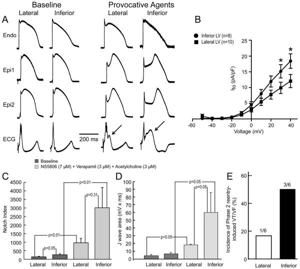
Figure 7.
Basis for greater vulnerability of inferior wall to early repolarization syndrome-mediated arrhythmia. Results shown are from left ventricular (LV) wedge preparations isolated from the inferior and lateral regions of the same hearts. A: Under baseline conditions, the action potential (AP) notch is more prominent in the epicardium (Epi) of the inferior wall as compared with the lateral wall and the response (accentuation of AP notch, increase in J wave) to a combination of NS5806 (7 μM) + Verapamil (3 μM) + Acetylcholine (ACh) (3 μM) is much more pronounced in the Epi of the inferior wall. APs shown are recorded from the lateral and the inferior part of the LV of the same dog. Arrows show increased J wave upon application of provocative agents. Endo = endocardial. B: Transient outward current density is more pronounced in Epi myocytes isolated from inferior vs. lateral wall. C: Notch index of APs recorded from the Epi surface of paired inferior vs. lateral wedge preparations isolated from the same heart under baseline conditions and after exposure to the same combination of provocative agents [NS5806 (7 μM) + Verapamil (3 μM) + ACh (3 μM)]. Results are mean ± S.E.M. D: J wave area recorded from paired inferior vs. lateral wedge preparations isolated from the same heart under baseline conditions and after exposure to the same combination of provocative agents [NS5806 (7 μM) + Verapamil (3 μM) + ACh (3 μM)]. Results are mean ± S.E.M. E: Incidence of phase 2 reentry –induced VT/VF after exposure of the lateral and inferior wedge preparations isolated from the same heart to the same combination of provocative agents.

