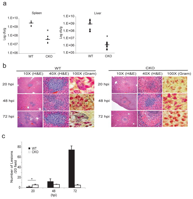
Figure 4. Myeloid-specific Blimp1 CKO mice are protected from L. monocytogenes infection.
a, WT or Blimp1 CKO mice were infected with 3×10⁴cfu of L. monocytogenes i.v. and bacterial loads were quantified in spleens and livers 48 hpi. Results from 3 different experiments are expressed as mean ± SEM. *p<0.001 (two-tailed Student’s t-test). b, Hematoxylin & Eosin staining (10X and 40X magnification) and Gram staining (100X magnification) of WT or CKO livers 20, 48 and 72 hpi by L. monocytogenes. The experiment shown is representative of 3 experiments conducted separately. c, Numbers of microabscesses per 20 fields of 3 different experiments were quantified and expressed as mean ± SEM. *p<0.05 (two-tailed Student’s t-test).

