Fig. 4.
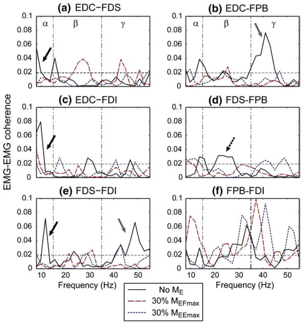
EMG–EMG coherence of the six muscle pairs for a single subject under three elbow moment conditions, i.e., no elbow moment, 30 % of maximum elbow flexion (30 % MEFmax), and 30 % of maximum elbow extension (30 % MEEmax): a EDC–FDS, b EDC–FPB, c EDC–FDI, d FDS–FPB, e FDS–FDI, and f FPB–FDI. The dotted horizontal lines denote the upper 95 % confidence limit. For this subject, a significant reduction in coherence values in the α-band of a subset of muscle pairs was apparent under elbow joint moment conditions (a, c, e solid black arrows). Coherence between the FDS–FPB pair was decreased mainly in the β-band (d dotted black arrow), and coherence between EDC–FPB and FDS–FDI pairs were affected in the γ band (b, e solid gray arrows). In contrast, for the intrinsic muscle pair (f FPB–FDI), the coherence value slightly increased under elbow joint moment conditions for this subject

