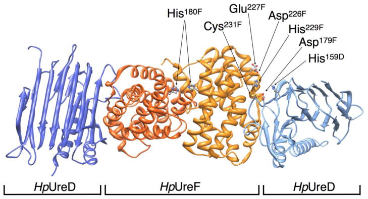
Figure 5.

Ribbon scheme of the H. pylori UreD-UreF complex (PDB code 3SF5), highlighting His180, His229, Cys231 on UreF and the nearby residues potentially involved in metal binding and trafficking. UreF monomers are depicted in dark and light orange, UreD monomers in dark and light blue.
