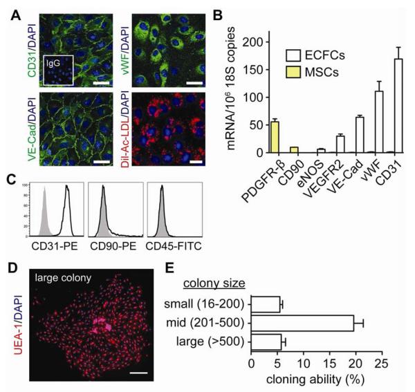
Figure 2. Assessment of ECFC phenotype.
A, ECFCs express CD31, vWF, VE-Cadherin, and uptake Dil-Ac-LDL. Cell nuclei were counterstained with DAPI (scale bar: 50 μm). B, Quantitative RT-PCR analyses of ECFCs for endothelial (CD31, vWF, VE-Cadherin, VEGFR2, eNOS) and mesenchymal (CD90, PDGFRβ) markers. C, Flow cytometric analysis of ECFCs for CD31, CD90, and CD45 (black line histograms). Isotype-matched controls are overlaid in solid gray histograms. D, Cloning-forming ability of ECFCs. The endothelial nature of the colonies was confirmed by binding of UEA-1 lectin (scale bar: 500 μm). E, Outgrown colonies were categorized by size. Bars represent mean±SE (n=6).

