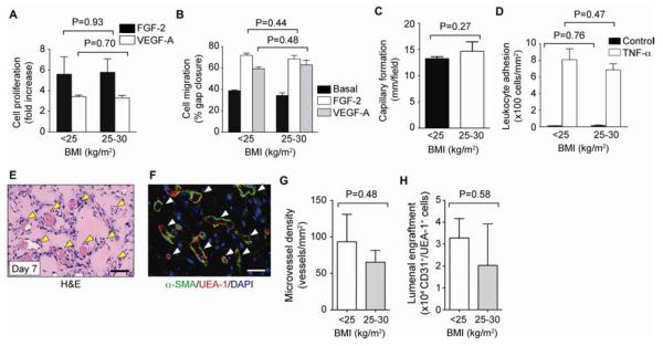
Figure 3. Assessment of ECFC function.
ECFCs were categorized into pre-pregnancy maternal BMI <25 kg/m2 and 25–30 kg/m2. A, Cell proliferation in response to VEFG-A (10 ng/mL) and FGF-2 (1 ng/mL). B, Cell migration in response to VEGF-A and FGF-2 expressed as percentage of gap closure. C, Capillary-like network formation on Matrigel expressed as total tube length per field. D, Adhesion of leukocytes onto ECFCs after stimulation with TNF-α. E, Vasculogenic properties of ECFCs compared in nude mice after subcutaneous implantation. H&E staining reveals numerous blood vessels (yellow arrowheads; scale bar: 100 μm). F, Human-specific lumens confirmed by staining with UEA-1 (red) and perivascular coverage indicated by α-SMA (green) (white arrowheads; scale bars: 50 μm). G, Microvessel density. H, ECFCs expressing UEA-1 and human CD31. Bars represent mean±SE (n=3 each group).

