Figure 7.
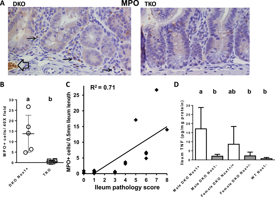
Analysis of MPO-positive infiltrating cells and TNF levels in the ileums of DKO and TKO mice. Panel A. MPO IHC of DKO and TKO ileum. The black arrows point to the positive stained monocytes in the submucosa of the crypt. The black open arrow points to a blood vessel and spuriously stained red blood cells. Panel B. A scatter plot of MPO+ cells analyzed in 50-day-old 5 DKO and 6 TKO males. The DKO group has higher number of MPO+ cells than TKO group (i.e. a>b; P=0.0048). Panel C. Correlation of MPO+ cells with ileum pathology scores. The correlation was made from all 11 mice in Panel B and five 28-day-old DKO mice. There is a positive correlation between MPO+ cells and pathology score with R2=0.71 (non-parametric). Panel D. A bar graph of TNF levels in mouse ileum at 50 days of age. The number of mice in each group is 3 male DKO, 8 male TKO, 4 female DKO, 6 female het-TKO and 8 WT Nox-mice of both genders. The error bars are SD. Groups with different letters are significantly different, where a>b. The groups share the same letter are not different, i.e. ab is not different from a or b.

