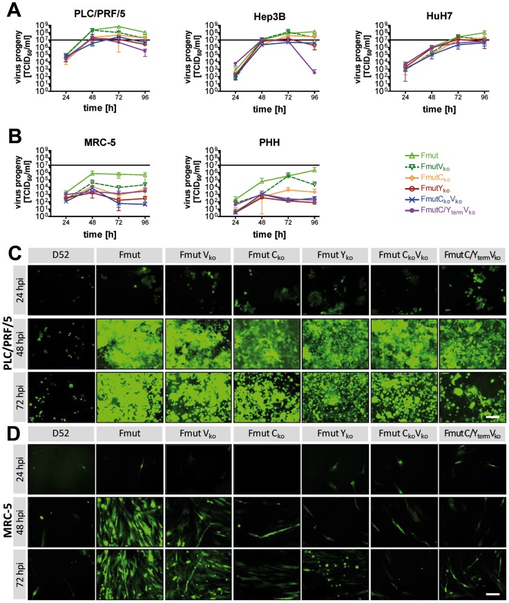
Figure 3. Growth kinetics and spreading of newly generated SeV variants in different cell types.
(A+B) Growth kinetics of six different recombinant SeV viruses over a 96×107 TCID50 is depicted. (A) Malignant human hepatoma cells PLC/PRF/5, Hep3B and HuH7. (B) Non-malignant MRC-5 fibroblasts and primary human hepatocytes (PHH) from three different donors. (C+D) Detection of EGFP reporter protein expression over a 72 h observation period by fluorescence microscopy as a surrogate marker for viral replication and spread to neighboring cells. Size bar: 200 µm. (C) Infection of PLC/PRF/5 hepatoma cells. (D) Infection of MRC-5 human fibroblast cells.

